Ever wondered how a simple ECG can reveal the secrets of your heart? Let’s dive into the world of leads on ECG and uncover what they really mean for heart health.
Understanding the Basics of Leads on ECG

Electrocardiography, commonly known as ECG or EKG, is a non-invasive diagnostic tool used to measure the electrical activity of the heart. At the heart of this technology—pun intended—are the leads on ecg. These leads are not physical wires leading to the heart but rather specific views or perspectives of the heart’s electrical activity captured from different angles on the body.
What Are Leads on ECG?
In ECG terminology, a ‘lead’ refers to a particular combination of electrodes placed on the skin that records the electrical potential difference between two or more points. Each lead provides a unique vantage point, allowing clinicians to observe how electrical impulses travel through the heart muscle during each heartbeat.
There are 12 standard leads in a conventional ECG: 6 limb leads and 6 precordial (chest) leads.These leads collectively create a 3D picture of cardiac electrical activity.The term ‘lead’ comes from the idea of ‘leading’ the signal from the body to the recording device.”The 12-lead ECG is one of the most valuable tools in cardiology—it’s fast, inexpensive, and incredibly informative.” — Dr.Eugene Braunwald, Harvard Medical SchoolHistorical Development of ECG LeadsThe concept of leads on ecg dates back to the early 20th century with Willem Einthoven, who invented the first practical ECG machine.
.He introduced the idea of using three limb leads—now known as Einthoven’s triangle (Leads I, II, and III)—to measure the heart’s electrical axis..
- Einthoven won the Nobel Prize in Physiology or Medicine in 1924 for his work.
- Later, Frank Wilson and colleagues developed the concept of ‘central terminal,’ enabling augmented limb leads (aVR, aVL, aVF).
- In the 1940s, the precordial leads (V1–V6) were introduced by Goldberger and others, completing the modern 12-lead system.
This evolution allowed for more precise localization of myocardial infarctions and arrhythmias, revolutionizing cardiac diagnostics.
The 12 Standard Leads on ECG Explained
To fully appreciate the diagnostic power of an ECG, it’s essential to understand the 12 standard leads on ecg. Each lead offers a distinct view of the heart, helping clinicians detect abnormalities in specific regions.
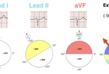
Limb Leads: The Foundation of ECG
The limb leads consist of three standard bipolar leads (I, II, III) and three augmented unipolar leads (aVR, aVL, aVF). They primarily assess the heart’s electrical activity in the frontal plane.
- Lead I: Measures voltage between the right and left arms.
- Lead II: From right arm to left leg—often used in monitoring due to its clear P wave.
- Lead III: Between left arm and left leg.
These form Einthoven’s triangle, a conceptual model where the heart sits at the center. The augmented leads (aVR, aVL, aVF) use a single positive electrode and a reference point derived from the other two limbs.
Lead II is often the default monitoring lead because it typically shows the most upright P waves, making atrial activity easy to track.
Precordial (Chest) Leads: Mapping the Horizontal Plane
The six precordial leads—V1 through V6—are placed across the chest and provide views of the heart in the horizontal (transverse) plane. These are crucial for detecting anterior, septal, and lateral wall issues.
- V1 and V2: Over the 4th intercostal space, right and left of the sternum—view the septum and right ventricle.
- V3 and V4: Transition zone; V4 is at the 5th intercostal space in the midclavicular line—views the anterior wall.
- V5 and V6: In the anterior and mid-axillary lines at the same level as V4—assess the lateral wall.
Additional leads like V7, V8, and V9 may be used in posterior MI cases, while V3R and V4R help evaluate right ventricular infarction.
How Leads on ECG Capture Heart Activity
The magic of leads on ecg lies in their ability to detect tiny electrical changes generated by cardiac depolarization and repolarization. When heart muscle cells contract, they produce electrical currents that spread through the body and can be picked up by surface electrodes.
The Electrical Axis and Lead Orientation
Each lead has a specific direction or vector. When the electrical impulse moves toward a positive electrode, it produces an upward deflection on the ECG; when it moves away, it creates a downward deflection.
- For example, in Lead II, the positive electrode is on the left leg, so any impulse traveling downward (like the normal P wave) appears upright.
- The QRS complex axis is calculated using the limb leads and should normally range from -30° to +90°.
- Left or right axis deviation can indicate conditions like left ventricular hypertrophy or right bundle branch block.
Understanding these vectors helps interpret which part of the heart is affected during ischemia or infarction.
Waveform Interpretation Across Leads
Different leads highlight different aspects of the ECG waveform: P wave (atrial depolarization), QRS complex (ventricular depolarization), and T wave (ventricular repolarization).
- In leads facing the left ventricle (like V5, V6, I, aVL), the QRS is usually predominantly positive.
- Lead aVR is unique—it typically shows inverted P waves, QRS complexes, and T waves because it ‘looks’ at the heart from the opposite direction.
- Abnormal Q waves in certain leads (e.g., II, III, aVF) may indicate prior inferior MI.
For instance, ST elevation in V1–V3 suggests anterior MI, while elevation in II, III, aVF points to inferior MI.
Clinical Significance of Leads on ECG
The real power of leads on ecg becomes evident in clinical settings where they guide life-saving decisions. From diagnosing heart attacks to identifying arrhythmias, each lead plays a critical role.
Diagnosing Myocardial Infarction Using ECG Leads
One of the most vital applications of leads on ecg is in the rapid diagnosis of acute myocardial infarction (MI). The location of ST-segment elevation or depression helps pinpoint the affected coronary artery.
- Anterior MI: ST elevation in V1–V4—often due to left anterior descending (LAD) artery occlusion.
- Inferior MI: ST elevation in II, III, aVF—usually from right coronary artery (RCA) blockage.
- Lateral MI: ST changes in I, aVL, V5, V6—linked to circumflex artery issues.
Posterior MI, though not directly visible, can be inferred from tall R waves and ST depression in V1–V3, confirmed with posterior leads (V7–V9).
The American Heart Association recommends obtaining a 12-lead ECG within 10 minutes of first medical contact for suspected MI. Source: American Heart Association
Detecting Arrhythmias Through Lead Analysis
Leads on ecg are indispensable in identifying various arrhythmias. For example:
- Atrial fibrillation shows irregularly irregular R-R intervals and absent P waves, best seen in Lead II and V1.
- Supraventricular tachycardia (SVT) may show narrow QRS complexes with retrograde P waves in inferior leads.
- Ventricular tachycardia often presents with wide QRS complexes and AV dissociation, visible across multiple leads.
Lead V1 is particularly useful for differentiating between supraventricular and ventricular tachycardias based on QRS morphology.
Common Misinterpretations of Leads on ECG
Despite their utility, leads on ecg are often misinterpreted, leading to incorrect diagnoses. Awareness of common pitfalls is crucial for accurate ECG reading.
Lead Misplacement Errors
Incorrect electrode placement is one of the most frequent errors affecting leads on ecg. Even small shifts can mimic pathology.
- Swapping left and right arm electrodes reverses Lead I and causes inversion in other limb leads.
- Placing chest leads too high or too low alters QRS amplitude and can mimic infarction patterns.
- Failure to place V4 at the 5th intercostal space can lead to misdiagnosis of anterior MI.
A study published in Journal of Electrocardiology found that up to 40% of ECGs have some degree of lead misplacement. Source: Journal of Electrocardiology
Normal Variants vs. Pathological Findings
Some ECG patterns that appear abnormal are actually normal variants, especially in young or athletic individuals.
- Early repolarization (ST elevation with notched J-point) is common in leads V4–V6 and is usually benign.
- Right bundle branch block (RBBB) pattern in V1 can be normal in healthy people.
- Q waves in III or aVL may be normal if no other signs of infarction are present.
Distinguishing these from true pathology requires correlation with clinical context and serial ECGs.
Advanced Applications of Leads on ECG
Beyond standard diagnostics, leads on ecg are being used in innovative ways to enhance cardiac care through technology and specialized monitoring.
Signal-Averaged ECG and Late Potentials
Signal-averaged ECG (SAECG) uses high-resolution analysis of multiple cardiac cycles to detect late potentials—small electrical signals after the QRS complex.
- Late potentials are associated with increased risk of ventricular tachycardia.
- SAECG analyzes filtered QRS duration, often using modified X, Y, Z leads orthogonal to standard leads.
- Used in patients with prior MI or cardiomyopathy to assess arrhythmia risk.
This technique enhances the predictive value of standard leads on ecg by detecting subtle conduction delays.
Body Surface Mapping and 80-Lead ECG Systems
Research systems use up to 80 electrodes to create detailed maps of cardiac electrical activity.
- Provides superior spatial resolution compared to 12-lead ECG.
- Helps localize arrhythmia origins and guide ablation therapy.
- Still primarily used in research but holds promise for clinical adoption.
These systems build upon the foundational principles of standard leads on ecg, offering a more granular view of heart function.
Future Trends in ECG Lead Technology
The future of leads on ecg is being shaped by wearable tech, AI integration, and remote monitoring, transforming how we capture and interpret cardiac data.
Wearable ECG Monitors and Reduced-Lead Systems
Devices like the Apple Watch, AliveCor KardiaMobile, and Zio Patch use fewer electrodes (1-3 leads) to provide continuous monitoring.
- KardiaMobile records a single-lead ECG comparable to Lead I.
- These devices are effective for detecting AFib and bradycardia but lack the comprehensive view of 12-lead ECG.
- They complement, rather than replace, standard leads on ecg in clinical settings.
Despite limitations, they increase accessibility and enable early detection of arrhythmias.
AI-Powered ECG Interpretation
Artificial intelligence is revolutionizing ECG analysis by enhancing accuracy and speed.
- AI algorithms can detect subtle patterns in leads on ecg that humans might miss.
- Google Health developed an AI model that predicts cardiovascular risk from retinal scans and ECG data.
- Some AI systems can estimate ejection fraction or detect hypertrophic cardiomyopathy from a standard 12-lead ECG.
A 2021 study in Nature Medicine showed AI could predict gender and age from ECG patterns, hinting at deeper physiological insights. Source: Nature Medicine
“AI won’t replace cardiologists, but it will augment their ability to interpret leads on ecg with unprecedented precision.” — Dr. Paul Friedman, Mayo Clinic
What do the 12 leads on ECG represent?
The 12 leads on ECG represent different electrical perspectives of the heart. Six limb leads (I, II, III, aVR, aVL, aVF) view the heart in the frontal plane, while six precordial leads (V1–V6) assess the horizontal plane, together providing a comprehensive 3D map of cardiac electrical activity.
How do leads on ECG help diagnose a heart attack?
Leads on ECG help diagnose a heart attack by showing characteristic changes like ST-segment elevation or depression in specific leads. For example, ST elevation in leads V1–V4 indicates anterior MI, while changes in II, III, aVF suggest inferior MI, helping locate the blocked coronary artery.
Can lead misplacement affect ECG results?
Yes, lead misplacement can significantly affect ECG results. Swapping limb electrodes or incorrect chest lead placement can mimic myocardial infarction, alter axis determination, or obscure arrhythmias, leading to misdiagnosis if not recognized.
What is the difference between bipolar and unipolar leads?
Bipolar leads (I, II, III) measure voltage between two electrodes, while unipolar leads (aVR, aVL, aVF, V1–V6) use one active electrode and a reference point. Augmented limb leads are unipolar but amplified to improve signal strength.
Are wearable ECG devices as accurate as standard 12-lead ECG?
Wearable ECG devices are useful for detecting certain arrhythmias like atrial fibrillation but are not as comprehensive as standard 12-lead ECG. They typically record 1-3 leads and cannot assess all heart regions, so they serve as screening tools rather than full diagnostic replacements.
Leads on ecg are far more than just lines on a graph—they are windows into the heart’s electrical soul. From Einthoven’s early discoveries to AI-driven diagnostics, these leads have evolved into a cornerstone of modern cardiology. Understanding their function, limitations, and clinical applications empowers both healthcare providers and patients. Whether diagnosing a life-threatening MI or monitoring a benign rhythm, the 12 leads on ECG remain an indispensable tool. As technology advances, the future promises even greater insights, making cardiac care more precise and accessible than ever before.
Further Reading: